Third Harmonic Generation Microscopy (THG)
In Third Harmonic Generation Imaging microscopy, the fluorescent dye is omitted and the third harmonic signal is generated from the sample itself. In a homogenous sample, the THG signal from above the focus cancels the signal generated below the focus due to phase matching. Thus, a third harmonic signal is only generated when the focus is close to a gradient in the refractive index. As a result, THG microscopy is particularly well-suited for 3D label-free imaging of transparent specimens where the membrane or other interface is of interest.
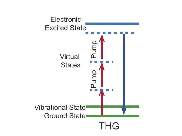
Figure 1. Energy level diagram for the THG imaging mechanism.
Third Harmonic Generation Bio-Imaging Examples
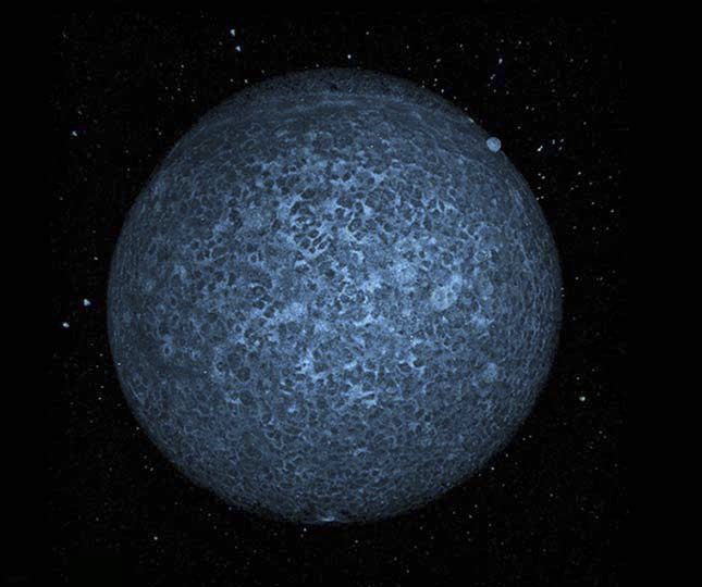
Figure 2. Zebrafish embryo (6 hours post fertilization) using label free THG at 1140 nm; imaged with InSight® DS+™ fs laser.
Courtesy of Dr. Nadine Peyrieras, CNRS, Gif sur Yvette, France
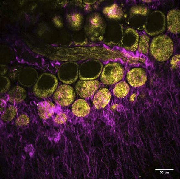
Figure 3. Mouse mammary gland, label free image of collagen (SHG, magenta) and adipocytes (THG, yellow), imaged with InSight DS+™.
Courtesy of Dr. Marie Irondelle, Institut Curie/CNRS, Paris, France
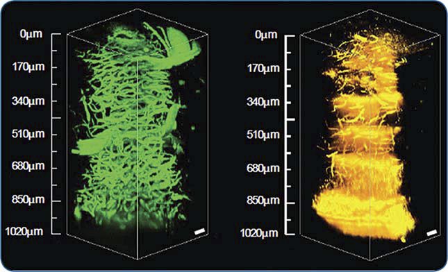
Figure 4. 3D images from a mouse brain cerebellum extending 1 mm deep into the tissue acquired via 3PF (left) and THG (right) microscopy at 1.3 μm using a Spirit laser with a Spirit-NOPA.
Courtesy of Chris Xu, with permission from SPIE Publications: Wang, et. al., “In vivo three-photon imaging of deep cerebellum,” Proc. SPIE: Multiphoton Microscopy in the Biomedical Sciences XVIII, vol. 10498, 2018.e