Stimulated Raman Scattering Microscopy (SRS)
In SRS, both the pump and Stokes frequencies are incident on the sample. If the frequency difference matches a molecular vibration of the sample, stimulated excitation of the vibrational transition occurs. SRS-based microscopy is a relatively recent development having been demonstrated in 2008 and gaining commercial viability only in recent years. Please see CARS and SRS Raman Microscopy for additional information.
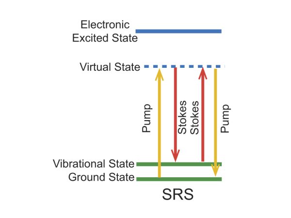
Figure 1. Energy level diagram for the SRS imaging mechanism.
Stimulated Raman Scattering Bio-Imaging Examples
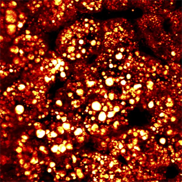
Figure 2. SRS image of fatty liver, pump 802 nm and Stokes at 1040 nm; acquired with InSight DS+.
Courtesy of Dr. Ji-XIn Cheng, Purdue University
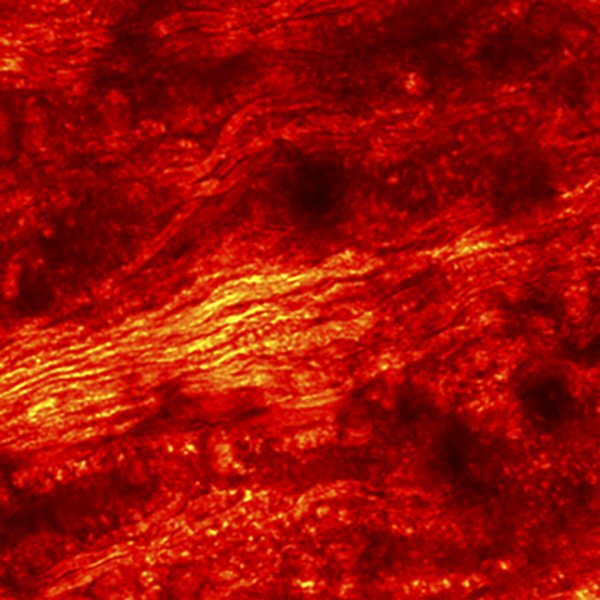
Figure 3. SRS image of spinal cord, pump 802 nm and Stokes at 1040 nm; acquired with InSight DS+.
Courtesy of Dr. Marc van Zandvoort, Maastrich University