Sum-Frequency Generation Microscopy (SFG)
In SFG microscopy, the fluorescent dye is omitted and the sum frequency signal is generated from the sample itself, similar to both SHG and THG microscopy. However, in contrast to these techniques where only a single wavelength is required, two different wavelengths are required for SFG. Consequently, this technique has similar requirements to CARS in that the pulses from the two lasers must arrive at the same location in the sample at the same time. Therefore, synchronized lasers are needed for SFG microscopy.
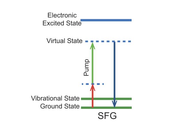
Figure 1. Energy level diagram for the SFG imaging mechanism.
An interesting application of multi-modal imaging is shown in Figure 2 where atherosclerotic plaque in an iliac artery was studied in using three different modalities: CARS, 2PF, and SFG. All three techniques work without labels and the sample was studied in vivo. The plaque shows up in the CARS image due to the strong signal from lipid bodies that are enriched in C-H bonds. The 2PF image (endogenous fluorophores generate two-photon excitation fluorescence or 2PF) also shows the lipid core while the SFG image reveals a lack of collagen fibrils in the same region.
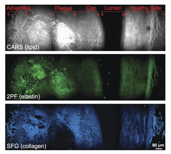
Figure 2. Cross-sectional view of an atherosclerotic plaque demonstrating multi-modal imaging.