Three-Photon Fluorescence Microscopy (3PF)
Three-photon fluorescence imaging (3PF) can provide a significant advantage over two-photon fluorescence imaging in strongly scattering samples such as a mouse brain. The fluorescence from three-photon excitation falls off as 1/z4, where z is the distance away from the focal plane, while the fluorescence from two-photon excitation falls off as 1/z2. As a result, three-photon excitation reduces the background from regions away from the focal plane and improves the signal-to-background ratio by orders of magnitude. Please see Three-Photon Fluorescence Microscopy for additional information.
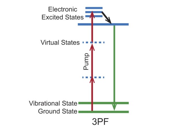
Figure 1. Energy level diagram for the 3PF imaging mechanism.
Three-photon Fluorescence Bio-Imaging Examples
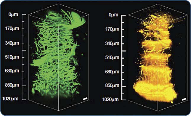
Figure 2. 3D images from a mouse brain cerebellum extending 1 mm deep into the tissue acquired via 3PF (left) and THG (right) microscopy at 1.3 μm using a Spirit laser with a Spirit-NOPA.
Courtesy of Chris Xu, with permission from SPIE Publications: Wang, et. al., “In vivo three-photon imaging of deep cerebellum,” Proc. SPIE: Multiphoton Microscopy in the Biomedical Sciences XVIII, vol. 10498, 2018.e
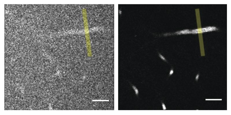
Figure 3. A comparison of images from 2PF (left, at 920 nm) and 3PF (right, at 1300 nm) microscopy of fluorescein-labeled blood vessels 650 µm deep in a mouse cerebellum. Scale bar, 50 µm.
Courtesy of SPIE Publications