Neuroscience
A single human brain contains more than 100 billion neurons. These neurons form a complex network with more than 100 trillion connections, or synapses, that send electrical impulses to control muscles and organs, and perform cognitive functions. An overarching objective in neuroscience is to develop an understanding of not only the physical structure, but more importantly, the functionality of the brain, especially the means of communication among neurons. Understanding these connections can reveal the fundamental science behind neural networks, which could lead to cures for diseases such as Parkinson's and Alzheimer's.
The discovery of Channelrhodopsin-2 (ChR2) spurred the first experiments of optogenetics and opened the possibilities for controlling neuronal activity using light. ChR2 is an opsin, a class of proteins comprised of different ion channels that can be opened and closed using light, i.e., photoactivation or photostimulation, and cause different signaling effects in the neurons that contain it. When an organism has been genetically engineered so its neurons contain opsins, neuronal activity can be triggered by illuminating the cell using bright light. Through photoactivation, the ChR2 channels pump cations into the cell, causing an increase of the voltage across the cell membrane (an action potential) and firing of the cell. This causes the release of neurotransmitters and signaling of neighboring neurons. This process is repeatable and can be done many times without causing damage to the cells. In order to activate a single cell, an ultrafast laser and a 2PF microscope are needed. By focusing the beam to a small spot around the neuron's membrane, sufficient power can be delivered to reach the threshold where the neuron will activate.
Courtesy of Sabine Scheibe, LMU Munich and Tilman Franke, FEI Munich GmbH
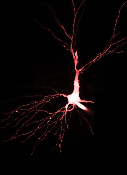
Courtesy of Gabriel Jones, Steven Petrou, University of Melbourne, Australia
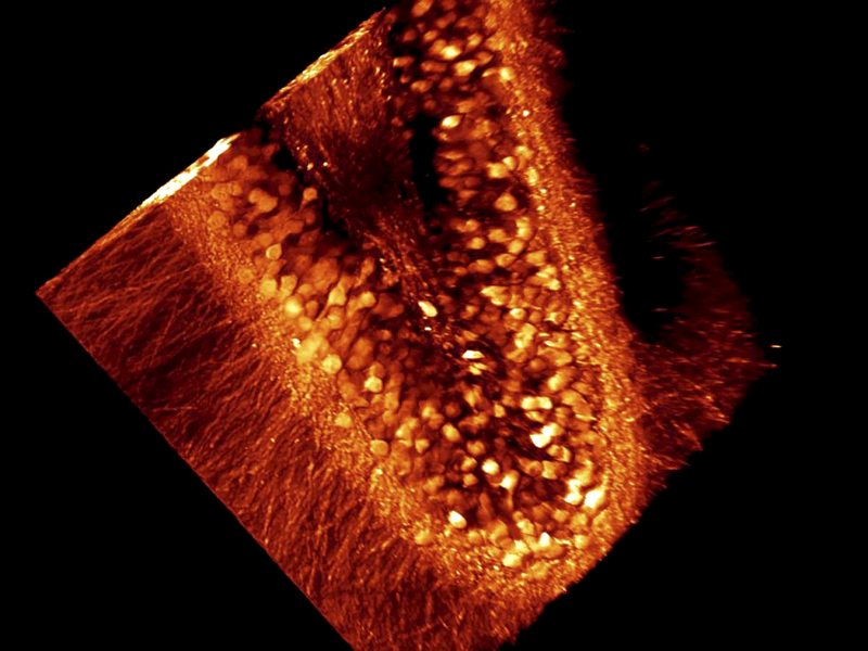
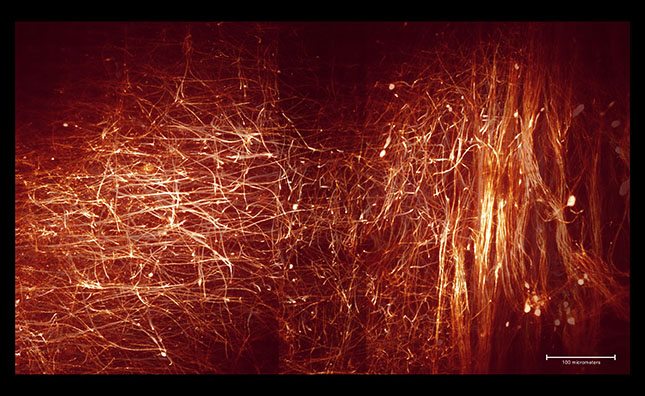
Courtesy of Karina Alvina, Albert Einstein College of Medicine
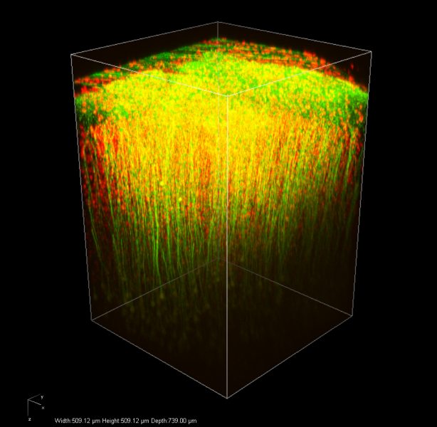
Courtesy of Naoki Honkura and Takeshi Imamura, Ehime University Graduate School of Medicine