Two-Photon Fluorescence Microscopy (2PF)
In 1990, workers at Cornell University demonstrated the use of two-photon excitation with laser scanning microscopy, denoted here as 2PF microscopy. In 2PF, two photons are simultaneously absorbed to cause a higher energy electronic transition in a fluorescent molecule as shown in Figure 1. Please see Two-Photon Fluorescence Microscopy for additional information.
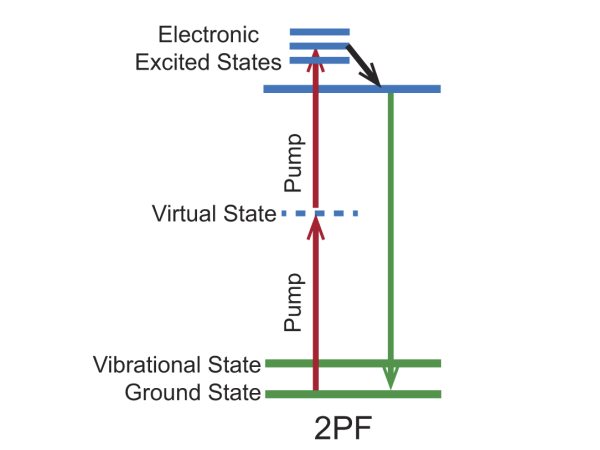
Figure 1. Energy level diagram for the 2PF imaging mechanism.
A schematic of a microscope system for 2PF is shown in Figure 2. A laser is focused to a tight spot in the specimen plane and scanned in a raster over the sample. When the laser focus overlaps with fluorescent molecules in the sample, fluorescence is generated selectively in the tiny focal volume and detected by photodetectors. The signal is spatially-mapped to generate individual pixels of an image by a data acquisition computer. The principal differences between confocal and 2PF microscopes are the laser and the fluorescence detection path. In 2PF microscopy, all fluorescent photons collected by the objective constitute useful signal as the detector pinhole is not required.
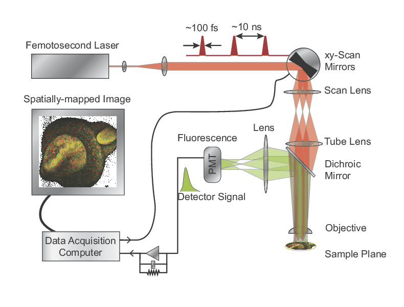
Figure 2. Schematic of a 2PF microscope.
Two-Photon Fluorescence Bio-Imaging Examples
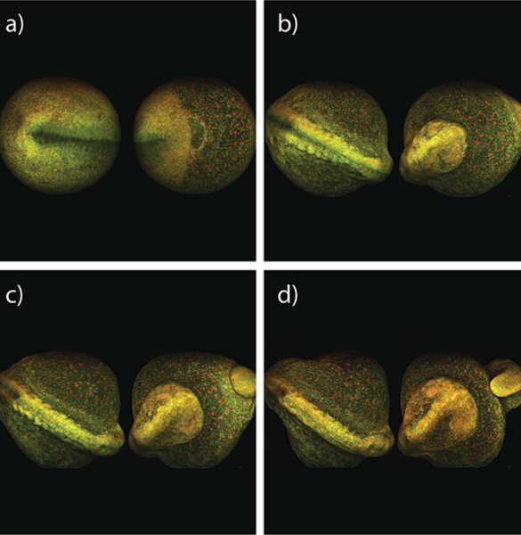
Figure 3. Maximum intensity projection of zebrafish embryo development, taken from both sides over 8 hours; 2PF imaged with InSight DS+.
Courtesy of Dr. Nadine Peyrieras, CNRS, Gif sur Yvette, France and LaVision Biotec
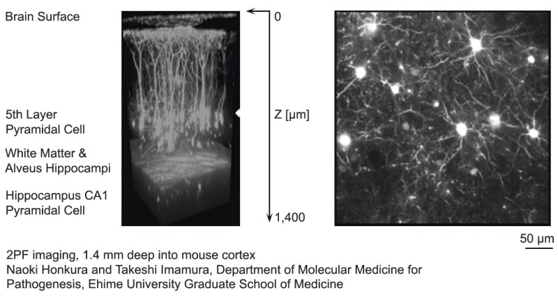
Figure 4. A 3D reconstructed image of a mouse cortex up to a depth of 1.4 mm using 2PF microscopy.
Courtesy of Naoki Honkhura and Takeshi Imamura, Ehime University Graduate School of Medicine
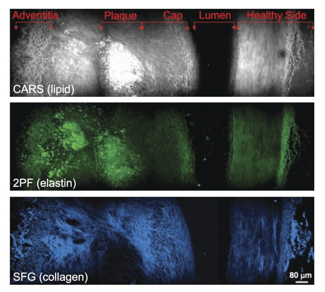
Figure 5. Cross-sectional view of an atherosclerotic plaque demonstrating multi-modal imaging.