受激拉曼光谱
在SRS 中,泵浦光和斯托克斯光都入射 到样品上。如果频率差与样品的分子振动相匹配,就会激发振动跃迁。基于SRS 的显微技术是一种相对较新的技 先进的生物成像 术,已在2008 年被证明,并在最近几年实现了商品化。
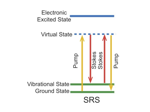
Figure 1. Energy level diagram for the SRS imaging mechanism.
Stimulated Raman Scattering Bio-Imaging Examples
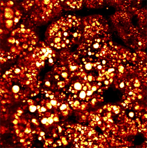
Figure 2. SRS image of fatty liver, pump 802 nm and Stokes at 1040 nm; acquired with InSight DS+.
Courtesy of Dr. Ji-XIn Cheng, Purdue University
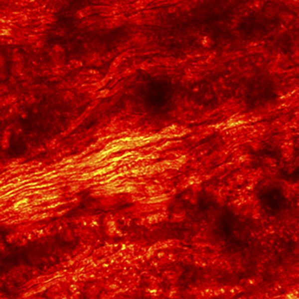
Figure 3. SRS image of spinal cord, pump 802 nm and Stokes at 1040 nm; acquired with InSight DS+.
Courtesy of Dr. Marc van Zandvoort, Maastrich University