相干反斯托克斯拉曼光谱
在CARS 显微镜中,泵浦光和斯托克斯光被紧密聚焦到样品中,通过扫描样品或激光束生成CARS 图像。除 了具有与2PF 和3PF 显微镜相同的3D 切片功能外,CARS 显微镜还有几个优点。CARS 显微镜允许无任何标记 的无损分子成像。这一优势对于脂质等小分子成像很重要,因为标记可能会显著影响其分子特性。来自分子振动 的相干放大产生了高方向性的输出,极大地方便了信号的采集。
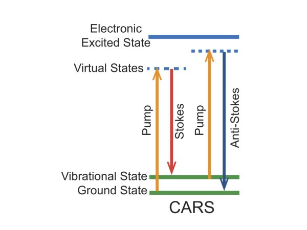
Figure 1. Energy level diagram for the CARS imaging mechanism.
Coherent anti-Stokes Raman Scattering Bio-Imaging Examples
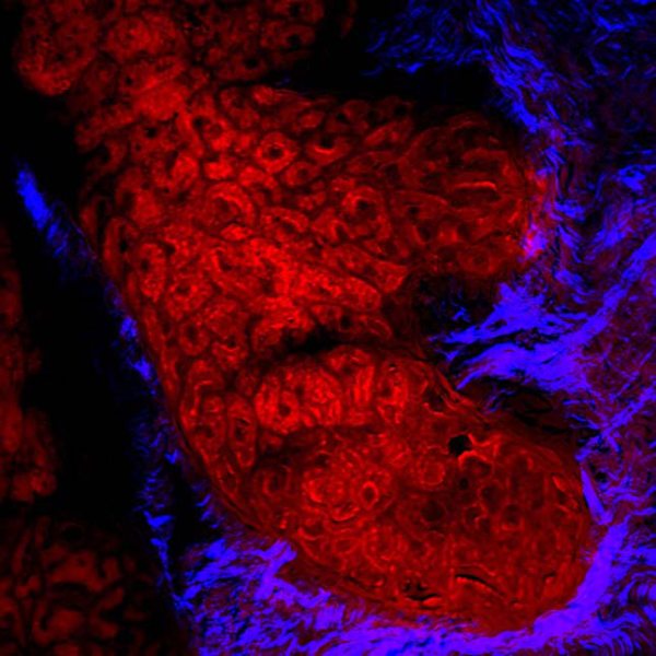
Figure 2. Human meibomian gland, CARS imaging the lipid rich meibocytes and SHG visualizing the surrounding collagen; acquired with InSight® DS+™.
Courtesy of Dr. Eric Potma, UC Irvine
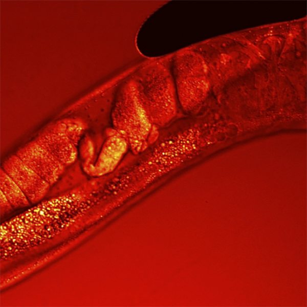
Figure 3. CARS Z-stack of C.Elegans worm, visualizing lipids 19.
Courtesy of Dr. Marc van Zandvoort, Maastrich University
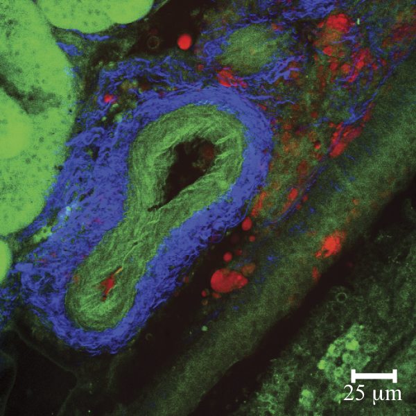
Figure 4. Multi-Modal images of mouse kidney featuring CARS (lipids, red) SHG (collagen, blue) and MPE (elastin autofluorescence, green); imaged with Mai Tai® HP plus Inspire™ OPO.
Courtesy of Dr. Eric Potma, UC Irvine, CA
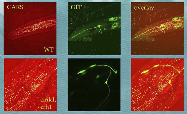
Figure 5. CARS and MPEF imaging of C. Elegans, neuron cells labelled with GFP, lipid droplets revealed with CARS.
Courtesy of Dr. Daewon Moon and Dr. Hyunmin Kim, DGIST Daegu Gyeongbuk Institute of Science and Technology (DGIST)
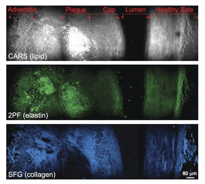
Figure 6. Cross-sectional view of an atherosclerotic plaque demonstrating multi-modal imaging.