三光子荧光显微镜
三光子荧光(3PF) 显微镜在强散射样品( 如小鼠大脑) 中比2PF 显微镜具有更明显的优势。三光子激发的荧 光衰减为1/z4,其中z 为与焦平面的距离,而双光子激发的荧光衰减为1/z2 。因此,三光子激发减少了远离 焦平面区域的背景,信号背景比提高了数个量级。图3 中将2PF 显微镜和3PF 显微镜进行了比较,说明了这种 背景的减少。
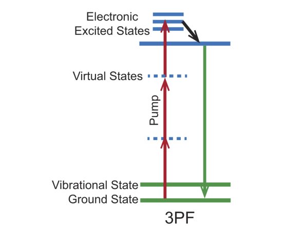
Figure 1. Energy level diagram for the 3PF imaging mechanism.
Three-photon Fluorescence Bio-Imaging Examples
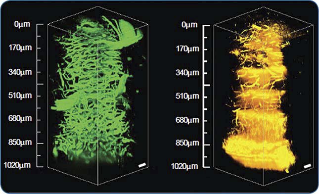
Figure 2. 3D images from a mouse brain cerebellum extending 1 mm deep into the tissue acquired via 3PF (left) and THG (right) microscopy at 1.3 μm using a Spirit laser with a Spirit-NOPA.
Courtesy of Chris Xu, with permission from SPIE Publications: Wang, et. al., “In vivo three-photon imaging of deep cerebellum,” Proc. SPIE: Multiphoton Microscopy in the Biomedical Sciences XVIII, vol. 10498, 2018.e
![2PF 显微镜( 左,920 nm) 和3PF 显微镜( 右,1300 nm) 荧光标记血管650 μm 深小鼠小脑图像的比较。这两幅图像 具有相似的信号强度,并在相同的对比度设置下显示。比例尺为50 μm。图片转载自文献[361],经过SPIE 出版社授权许可。](/mam/celum/celum_assets/Figure_304-Photonics_Handbook_800w.jpg)
Figure 3. 2PF 显微镜( 左,920 nm) 和3PF 显微镜( 右,1300 nm) 荧光标记血管650 μm 深小鼠小脑图像的比较。这两幅图像 具有相似的信号强度,并在相同的对比度设置下显示。比例尺为50 μm。图片转载自文献[361],经过SPIE 出版社授权许可。
Courtesy of SPIE Publications