双光子荧光显微镜
传统的宽场显微镜提供微米量级的横向分辨率。相反,由于来自样品内部不同深度的信号会导致样品的原点 模糊,因此对样品深度的分辨率( 有时标记为Z 方向) 可能会低得多。在共聚焦激光扫描显微镜中,聚焦激光产生 的荧光在针孔或共焦孔径上成像,在到达探测器之前,针孔或共焦孔径会阻止非焦平面上产生的荧光。然后测量 荧光强度,当光束水平方向扫描样品时,形成一个二维图像。最后,样品按Z 方向顺序移动,允许以接近衍射极 限的空间分辨率构建三维图像。这种共聚焦激光扫描显微镜技术可以追溯到1969 年。
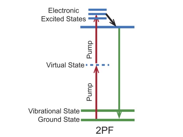
Figure 1. 荧光染料的2PF 能级图。
2PF 显微镜系统示意图如图2 所示。激光被聚焦到试样平面的一个小点上,然后按栅格扫描试样。当激光 聚焦与样品中的荧光分子重叠时,在微小的聚焦体积内选择性地产生荧光,并被光电探测器检测。通过数据采集 计算机对信号进行空间映射,生成图像的各个像素。共焦显微镜和2PF 显微镜的主要区别在于激光和荧光的检测 路径。在2PF 显微镜中,由于不需要探测针孔,目标所收集的所有荧光光子都是有用的信号。
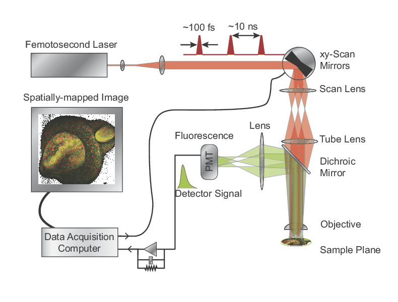
Figure 2. 2PF 显微镜示意图.
Two-Photon Fluorescence Bio-Imaging Examples
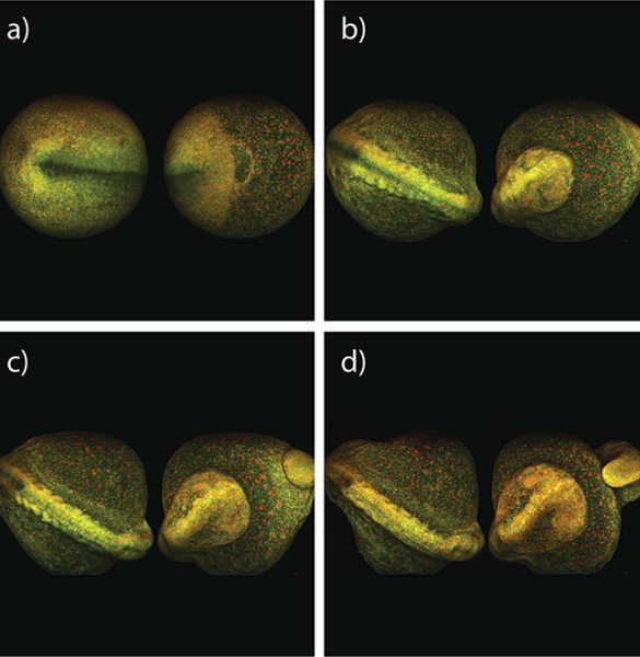
Figure 3. Maximum intensity projection of zebrafish embryo development, taken from both sides over 8 hours; 2PF imaged with InSight DS+.
Courtesy of Dr. Nadine Peyrieras, CNRS, Gif sur Yvette, France and LaVision Biotec
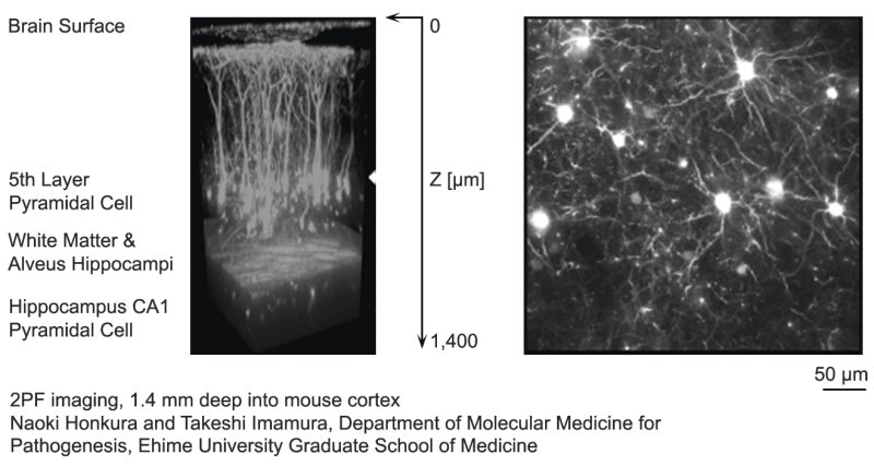
Figure 4. A 3D reconstructed image of a mouse cortex up to a depth of 1.4 mm using 2PF microscopy.
Courtesy of Naoki Honkhura and Takeshi Imamura, Ehime University Graduate School of Medicine
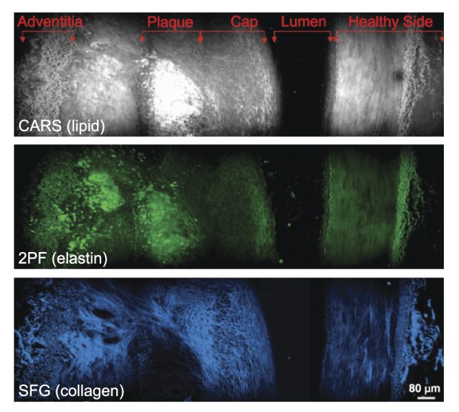
Figure 5. Cross-sectional view of an atherosclerotic plaque demonstrating multi-modal imaging.