Second Harmonic Generation Microscopy (SHG)
Second Harmonic Generation Imaging (SHG) is a second order coherent process where two lower energy photons are up-converted to exactly twice the incident frequency of an excitation laser. It has uses in many Bio-imaging applications.
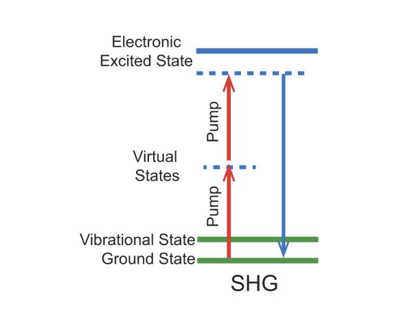
Figure 1. Energy level diagram for the SGH imaging mechanism.
Second Harmonic Generation Bio-Imaging Examples
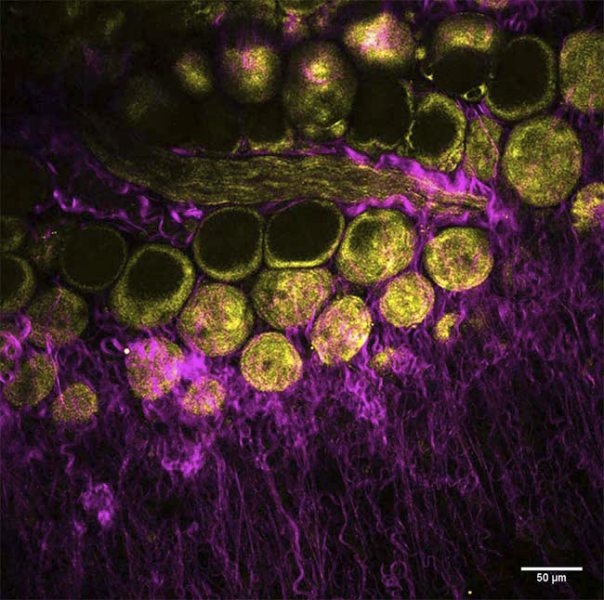
Figure 2. Mouse mammary gland, label free image of collagen (SHG, magenta) and adipocytes (THG, yellow), imaged with InSight DS+™.
Courtesy of Dr. Marie Irondelle, Institut Curie/CNRS, Paris, France
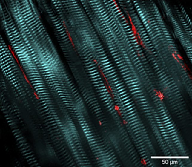
Figure 3. Mouse blood vessel wall, SHG and multiphoton excited fluorescence imaging with HighQ-2™ fs laser.
Courtesy of Dr. Marc van Zandvoort, Maastrich University
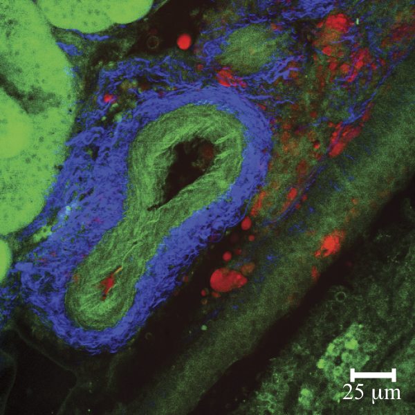
Figure 4. Multi-Modal images of mouse kidney featuring CARS (lipids, red) SHG (collagen, blue) and MPE (elastin autofluorescence, green); imaged with Mai Tai® HP plus Inspire™ OPO.
Courtesy of Dr. Eric Potma, UC Irvine, CA
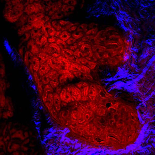
Figure 5. Human meibomian gland, CARS imaging the lipid rich meibocytes and SHG visualizing the surrounding collagen; acquired with InSight® DS+™.
Courtesy of Dr. Eric Potma, UC Irvine