Coherent anti-Stokes Raman Scattering Microscopy (CARS)
In CARS imaging, the pump and Stokes beams are tightly focused into the sample and a CARS image is generated by scanning either the sample or the laser beams. Besides possessing the same 3D sectioning capability afforded to 2PF and 3PF microscopy, CARS microscopy also permits non-destructive molecular imaging without any labeling. Please see CARS and SRS Raman Microscopy for additional information.
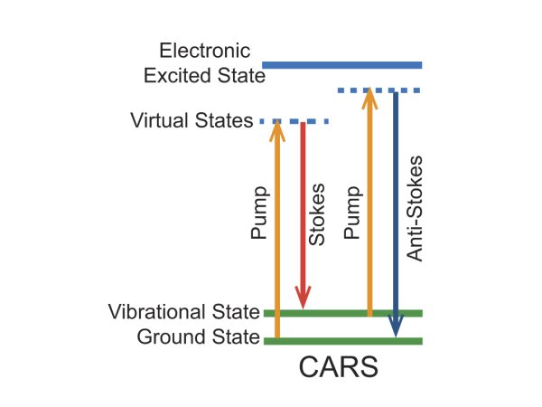
Figure 1. Energy level diagram for the CARS imaging mechanism.
Coherent anti-Stokes Raman Scattering Bio-Imaging Examples
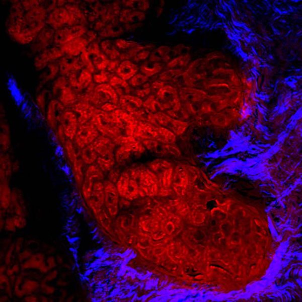
Figure 2. Human meibomian gland, CARS imaging the lipid rich meibocytes and SHG visualizing the surrounding collagen; acquired with InSight® DS+™.
Courtesy of Dr. Eric Potma, UC Irvine
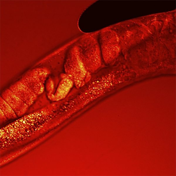
Figure 3. CARS Z-stack of C.Elegans worm, visualizing lipids 19.
Courtesy of Dr. Marc van Zandvoort, Maastrich University
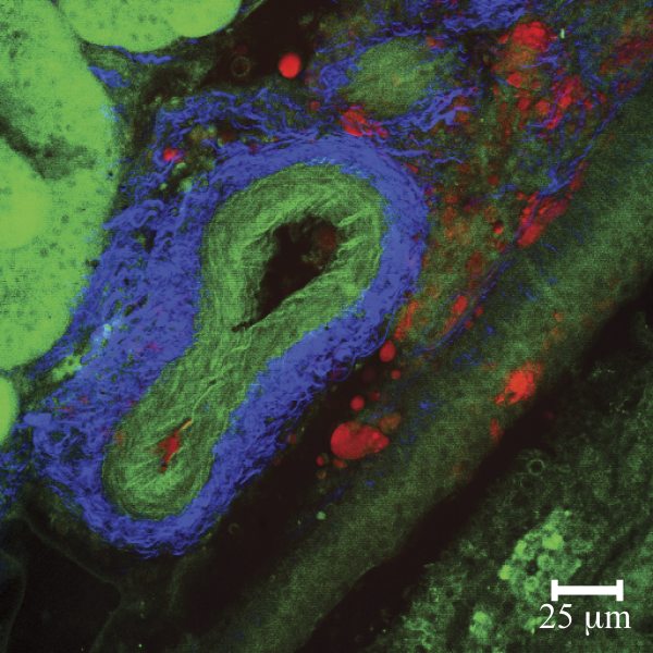
Figure 4. Multi-Modal images of mouse kidney featuring CARS (lipids, red) SHG (collagen, blue) and MPE (elastin autofluorescence, green); imaged with Mai Tai® HP plus Inspire™ OPO.
Courtesy of Dr. Eric Potma, UC Irvine, CA
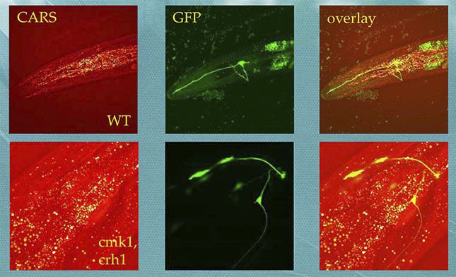
Figure 5. CARS and MPEF imaging of C. Elegans, neuron cells labelled with GFP, lipid droplets revealed with CARS.
Courtesy of Dr. Daewon Moon and Dr. Hyunmin Kim, DGIST Daegu Gyeongbuk Institute of Science and Technology (DGIST)
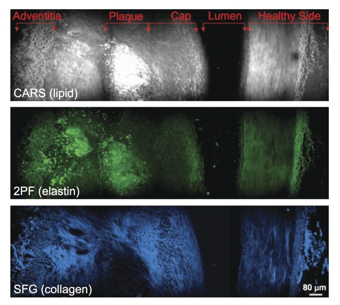
Figure 6. Cross-sectional view of an atherosclerotic plaque demonstrating multi-modal imaging.
 Ultra-High Velocity